Miracle protein ( Bone Morphogenetic Protein ) transforms cleft surgery at Balaji Dental & Craniofacial Hospital
Approximately 1 in every 500-700 children is born with a cleft lip, cleft palate or both, worldwide. Cleft lip and cleft palate are birth defects that affect the upper lip and roof of the mouth. They happen when the tissues that form the roof of the mouth and upper lip don’t join properly before birth resulting in a gap. This can affect the way the child’s face looks. It can also lead to difficulties in breathing, eating, talking, ear infections and psychological problems.
born with a cleft palate have a defect in the upper alveolar (tooth bearing) region. Conventionally, this has been treated with bone graft surgery, harvesting bone from the patient’s rib or hip. A potential downside of this is that it involves an additional surgery incurring undesirable scars and increases blood loss, hospital stay, recovery time and pain. It also may not be a viable option in instances where the patient’s overall bone quality is poor or when a large volume of bone is required.
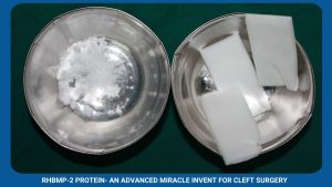
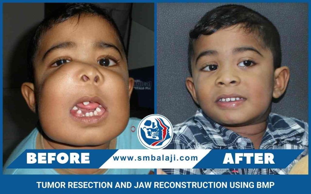
A recent and revolutionary invent, rhBMP-2 (recombinant human Bone Morphogenetic Protein), is successfully used to treat jaw bone defects at Balaji Dental and Craniofacial Hospital. This is a genetically engineered version of a protein that is normally found in the body. Dr. S.M. Balaji is credited to be the first to introduce this novel technology in India.
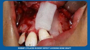
The inclusion of rhBMP-2 at the bone deficient site stimulates the body’s stem cells to rapidly produce new bone and promotes growth ultimately healing the bone defect. The need for additional bone graft surgery, unaesthetic scars, pain or discomfort at the donor site is overcome. rhBMP-2 now ensures superior results to the conventional alveolar bone grafting techniques by allowing alveolar cleft closure at a younger age of 3 to 4 years rather than waiting till 7 to 8 years. This technology has greatly improved the quality of life of innumerable people, more importantly children, afflicted with jaw bone defects.
If the upper jaw is comparatively bigger than the lower jaw, it may lead to a “gummy” smile. Excess gum tissue may be visible which may deflect from the beauty of the smile. This disparity in the size and shape of the jaw bones can be corrected by Orthognathic surgery. The jaw size is decreased and the jaw is placed in a normal position drastically improving smile and appearance.
The shape of the nose is influenced by the shape of the upper jaw. Sometimes, the sharpness of the nose is not apparent because the upper jaw is too big and protruding. In such cases, correcting the shape, size and position of the upper jaw also results in giving the nose a sharp pristine look. More severe variations in the shape of the nose can be corrected along with jaw correction in the same surgical procedure.
Before deciding the line of treatment, a detailed assessment and analysis of the face & facial bones needs to be done. Many a times, there is often a short period of orthodontic treatment done for subtle and fine detailing of the tooth positions.
The advantage of orthognathic surgery is that immediate results are seen. Also since these surgeries are approached from inside the mouth there are no visible external scars.
Jaw correction surgery brings the teeth and jaws into proper position for better health and appearance. For some patients, the surgery can dramatically enhance appearance and self esteem. Such surgeries hold the key for youngsters who go through a lot of psychological stress over their facial appearance.
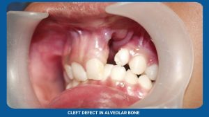
A 14-year-old boy from Sri Lanka, with unilateral cleft lip and palate, was brought to our hospital by his parents for surgical correction of alveolar cleft (cleft in the teeth-bearing bone of upper jaw). Previously he was operated for cleft lip & cleft palate elsewhere.
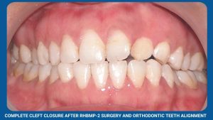
Maxillofacial Surgeon Dr. S.M. Balaji performed the surgical correction of the alveolar cleft using the advanced rhBMP-2 (recombinant human Bone Morphogenetic Protein). This miracle protein stimulates the body’s own stem cells to form new bone at the site where it’s placed ultimately healing the bone defect. Following this, normal teeth eruption and teeth alignment can be achieved. The wonder of using this advanced technique is that additional surgery of bone grafting from the rib or hip is avoided and there is no scarring.

