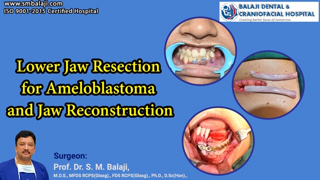
Lower Jaw Resection for Ameloblastoma and Jaw Reconstruction
Pathogenesis of Ameloblastoma and Treatment Modes
An ameloblastoma is a benign odontogenic tumor that most often arises in the lower jaw. The most common region is the area near the molar teeth. It is often aggressive in nature and causes extensive destruction of the jaw bone.
Successful Surgical Treatment of Ameloblastoma with Segmental Resection
Segmental jaw resection is the most common surgical mode of treatment of ameloblastoma. The section of affected jaw along with overlying teeth is removed. This results in a large bony defect, which leads to cosmetic and functional compromise.
Common Modes of Diagnosis of Ameloblastoma of the Jaw
Diagnosis is most commonly through biopsy and imaging studies. It appears as a radiolucent lesion, which is well defined and with scalloped borders. The scalloped borders are caused by the multilocular presentation of the tumor.
Histopathological Genesis of the Tumor in the Mandible
Ameloblastoma of the jaw arises from the cells that are the precursor to enamel. Occurrence in children can lead to delayed eruption of teeth. Adults often experience loosening of teeth in the region and difficulty with speech.
Extensive Destruction of Bony Structure of the Mandible
Patients who develop ameloblastoma of the jaw require jaw reconstruction surgery. Jaw bone grafting needs to be performed using rib bone grafts. A time period of six months is allowed to elapse after jaw bone grafting. This allows adequate consolidation of the graft bone.
Surgical Specialty Dealing with the Treatment of Ameloblastoma
Treatment of ameloblastoma of the jaw is performed by maxillofacial surgeons. This is achieved by segmental resection of the affected segment of the jaw. This is followed by jaw reconstruction surgery.
Patients who develop ameloblastoma of the jaw fully recover from their condition. Jaw reconstruction surgery is followed by placement of dental implants. Shade matched ceramic crowns are placed after osseointegration of the implants.
Treatment plan with this surgical procedure includes long term correct jaw relationship. A CT scan is obtained before this corrective jaw surgery. All possibilities for side effects are studies to ensure that they do not arise.
The soft tissues also play an important role in determining the shape of the face. Relationship between the upper and lower jaw is studied in detail. The bone structure is also analyzed. This will help chose the choice of bone graft material.
Bone graft procedures are explained in detail to the patient. Care is also taken to avoid complications such as a postoperative open bite. The gum tissue is also analyzed for postoperative drape.
These procedures are always performed under general anesthesia. They also often require orthodontic treatment for correction of tooth alignment. Patients with obstructive sleep apnea have a characteristic profile with receding chins.
Successful Rehabilitation of Patients with Full Return to Normalcy
Our hospital is a superspecialty oral and craniomaxillofacial surgery hospital. Located in Chennai, India, it has won international acclaim for its surgical services. We address all conditions related to the craniofacial complex.
Dr. SM Balaji of Balaji Dental, Craniofacial Hospital & Research Institute is a world renowned craniofacial surgeon. He has over 30 years of experience in handling complex cases.
Delivery of healthcare is holistic in nature in our hospital. We believe in the overall wellbeing of the patient. Education is imparted to patients regarding the impact of oral health on overall health.
Our approach to treatment came to the fore during the recent COVID-19 pandemic. We have always placed the greatest importance to overall patient wellbeing. Modules were developed to educate patients on reducing their risk to getting infected.
Stress was laid on the wear masks, wash hands and maintain distance protocol. We have always had the latest infrastructure regarding asepsis and infection control. Staff were regularly updated on information developed by leading scientists.
We were also amongst the first to commence surgeries in our nation. Diligent attention was paid to ensure that patient safety was always ensured. Our untiring efforts won recognition from many quarters across the nation.
World Class Amenities at our Superspecialty Surgical Center
We have been rendering excellence in surgical care for over three decades now. Our hospital has two state of the art operation theaters with the latest equipment. We also have 24 well-appointed rooms for inpatient admissions.
Continuous Medical Education Programs for Staff Members
Our medical and paramedical staff are up to date with the latest surgical developments. We are credited with many surgical innovations in the field of craniomaxillofacial surgery. Many Western surgeons apply for viewership in our operation theaters.
We keep abreast with the latest developments in the surgical sciences. Jaw bone grafting is exclusively performed using rib grafts. Precise jaw bone grafting requires a high degree of skill and experience. Rib bone grafts are the gold standard for use in jaw bone grafts.
Patient Notices Insidious Development of Lower Jaw Swelling
This young woman had noticed a swelling involving her left lower jaw for a few months now. She had noticed that it was also painful when chewing and biting. This had begun to alarm her as it kept increasing in size.
Diagnosis of Ameloblastoma through Imaging Studies and Biopsy
Imaging studies and biopsies were obtained upon presentation at our hospital. This had returned with a diagnosis of ameloblastoma involving the left lower jaw. Treatment planning for jaw reconstruction surgery was explained to the patient.
Harvesting Rib Bone Graft for Reconstruction for Jaw Bone
Surgery commenced with harvesting of a rib bone graft. This was followed by segmental resection of the lower jaw ameloblastoma. Titanium reconstruction plate and the rib graft was used to reconstruct the bony defect.
Successful Rehabilitation of Patient with Dental Implants and Crowns
The patient was instructed to return in six months for dental implant placement. This would allow for consolidation of the grafts with surrounding alveolar bone. Another six months would be allowed for osseointegration of the implants. This will be followed by dental crown fixation.
Patient’s Reflection
In her own words, the patient shares her reflections on the surgical experience, emphasizing the support received from the medical team, the postoperative healing process, and the gradual restoration of normalcy. Her testimony serves as a beacon of hope for others facing similar challenges.
“I never imagined facing such a journey, but the team at Balaji Dental and Craniofacial Hospital became my pillars of strength. From the initial diagnosis to the surgery and the follow-up care, their commitment to my well-being was unwavering. Today, I stand on the other side of adversity, grateful for the transformation that jaw reconstruction has brought to my life. I share my story with the hope that it inspires others to find courage and seek the right care for their journey to recovery.”


