Craniofacial Deformities
Craniofacial Deformities: Embracing Diversity in Facial Structures
Craniofacial deformities encompass a wide range of irregularities that impact the shape and structure of the head and face. These anomalies may either be present from birth (congenital) or develop later in life due to factors such as injury, disease, or other influences. Their effects can vary from purely aesthetic concerns to more significant impacts on function and overall quality of life.
Varieties of Craniofacial Deformities:
Cleft lip and palate: A condition involving the separation of the lip and/or palate, influencing speech, feeding, and facial aesthetics.
Craniosynostosis: The premature fusion of skull bones, resulting in abnormalities in head shape.
Hemifacial microsomia: Underdevelopment of facial bones on one side.
Microcephaly: Abnormally small head size.
Macrocephaly: Abnormally large head size.
Mandibular deficiency: Underdeveloped jawbone.
Apert syndrome: A genetic condition affecting the hands, feet, and skull bones.
Causes of Craniofacial Deformities:
Genetics: Some deformities have a hereditary basis, while others may occur due to spontaneous mutations.
Environmental factors: Certain medications, infections, and environmental exposures during pregnancy can elevate the risk of craniofacial abnormalities.
Trauma: Injuries to the head and face can lead to bone fractures and deformities.
Tumor growth: Tumors in the head and neck region can impact bone growth and development.
Treatment Approaches:
The optimal treatment for craniofacial deformities depends on the specific type and severity of the condition. Treatment modalities may include:
Surgery: Corrects bone defects, enhances facial symmetry, and restores function.
Dental braces and orthodontics: Address jaw alignment and bite problems.
Speech therapy: Assists individuals with cleft lip and palate in developing proper speech patterns.
Prosthetics: Utilizes devices like facial implants or cranial helmets to enhance appearance.
Hypertelorism Unveiled: Exploring the World of Widely Spaced Eyes
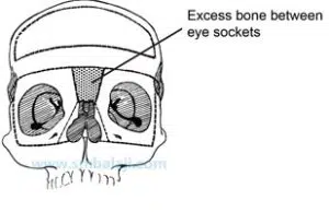
For a normal looking face, the distance between the eyes should be roughly 30 to 35 mm in children. Even slight increase makes a huge difference in appearance. Orbital Hypertelorism is the condition that refers to an abnormally increased distance between the two eyes measuring more than 35 mm in children. In this condition, the eyes are so wide set that the nose appears broad, almost animal like and flat. All the structures that normally occur between the eyes are displaced. Sometimes even the brain is positioned lower, and found hanging between the eyes. This condition occurs since birth and can occur alone or as manifestation of other birth deformities.
Have you ever observed someone with eyes that seem unusually far apart? This distinct feature characterizes hypertelorism, a medical term denoting an increased distance between the eye sockets (orbits). Whether subtle or striking, hypertelorism prompts inquiries about its origins, consequences, and potential remedies.
Unpacking Hypertelorism:
Distance Matters: Hypertelorism manifests as a greater-than-expected gap between the inner corners of the eyes and the pupils, influencing both facial aesthetics and, at times, vision.
Beyond Aesthetics: Frequently, hypertelorism serves as a symptom of an underlying condition, spanning genetic syndromes like Edwards syndrome to craniofacial disorders such as Apert syndrome or Crouzon syndrome. In rare instances, it may manifest independently, termed “isolated hypertelorism.”
Diagnosis and Management: Identifying hypertelorism typically involves a physical examination and, if necessary, imaging tests like X-rays or CT scans to pinpoint any root causes. Treatment approaches hinge on the specific cause and severity, with surgery being a potential option in certain cases to enhance facial aesthetics and eye alignment—though thorough evaluation is imperative.
CASH I : Complex eye, nose and skull position correction surgery performed for 9-year-old boy

The Challenge Correction of this kind of deformity is routinely carried out by Dr. S.M. Balaji though each case is different and the techniques used for the treatment need to be customized and modified. The deformity can be so severe that such children are isolated and live as recluses and therefore early correction of the deformity is mandatory. But attempting such a complex brain and eye repositioning surgery at such an early age is a colossal challenge. The total correction needs multiple steps but the primary and first stage of bringing the eyeballs to the normal position without any change in vision is the trickiest and challenging.
Improvisation
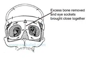
The technique of moving the eyeballs along with the sockets is called facial bipartition surgery. Dr. Balaji, a pioneer of such surgeries in India says every case poses a different challenge. In this particular case, there was a defect in the floor of the skull holding the brain and therefore extreme care needed to be taken to preserve unprotected brain tissue. Moreover, the child had very small eyes and the upper half of the face was flattened completely due to the lack of growth around the eyes.
The cheekbones were diminutive and set behind. Due to the combined presence of two different deformities, each requiring two different types of surgeries, Dr. Balaji decided to combine two techniques and decided to advance the face while bringing the eyes together. He also modified the technique further to prevent any scarring to the eyelids – by doing the entire surgical cuts through the mouth. This technique was very successful in bringing fullness to the child’s face while setting his eyes in the right place.
Case Details
A 9-year-old Master Suhail Khan from Cuddappah, Andhra Pradesh, was born with a facial deformity which made his look “different”. He had abnormally wide set eyes, a flat facial appearance and a deformed cleft nose. The distance between his eyes was 50 mm. His very wide set eyes were affecting his normal vision (like bird’s vision). He was referred to Balaji Dental & Craniofacial Hospital for specialized face surgery.
Dr. S.M. Balaji readily took up the challenge to help out the child and his parents in distress. A CT scan taken to gauge the extent of the deformity showed very widely set eye sockets, flattened cheek bones, nasal bone deformity but normal alignment of teeth
CASH II : Cranial synostosis with Plagiocephaly and hypertelorism

Another such similar surgery performed for a girl An eight year old girl with a similar birth defect was referred from Seychelles to Balaji Dental & Craniofacial Hospital. She was born with cranial synostosis with Plagiocephaly (flattening of cranial bones) and hypertelorism (far apart placed eyes “birds eye” and absence of binocular vision – like humans).
In a complex surgery, her skull bones were exposed, by making a 360° bony cut her eye socket bones were brought closer and fixed using bone plates. Now her eyeballs are in a more normal position. She will need nose correction surgery after about 3 to 4 years.


