Facial asymmetry correction- condylar hyperplasia already operated but failed, maxillary shortening, buccal fat pad transfer, malar and mandibular body augmentation surgery
Patient with failed surgery elsewhere presents for correction The patient is a young woman with failed condylar hyperplasia surgery performed elsewhere. She presented to our hospital for correctional surgery. Dr SM Balaji examined the patient and ordered detailed studies for the patient. Rib graft and buccal fat pad graft obtained from the patient Under general anesthesia, a rib graft was first harvested from the patient. Valsalva maneuver demonstrated a patent thoracic cavity. The incision was then closed with sutures. This was next followed by harvesting of a buccal fat pad graft from the right cheek. Augmentation of the mandibular body done with grafts A left sided maxillary vestibular incision was next made. The bone grafts were then shaped and fixed with screws in this region. Attention was next turned to the mandible. A mucogingivoperiosteal flap was then raised. The bone grafts were then screwed in place in the molar region. This led to adequate augmentation of the body of the mandible. Maxillary repositioning and buccal fat pad transfer performed Attention was next turned to the anterior maxillary region. An osteotomy was then performed and the maxilla repositioned with four holed plates. This was then followed by transfer of the buccal fat pad graft to the left cheek. All incisions were then closed with sutures. The patient expressed complete satisfaction with the results of the surgery before discharge. Surgery Video
Large dentigerous cyst of maxilla enucleated. Root canal treated teeth saved and defect filled with rib graft
Young boy with dentigerous cyst presents with nonvital teeth The patient is a young boy who presented with a swelling in the left anterior maxilla. Dr SM Balaji examined the patient. The teeth in the area of the swelling were nonvital. Radiograph revealed the presence of a large dentigerous cyst in that region. Treatment planning was for surgical excision of the cyst. Dentigerous cyst enucleated and teeth saved A mucogingivoperiosteal flap was first raised in the anterior maxillary region. There was a supernumerary tooth present within the dentigerous cyst. The dentigerous cyst was then enucleated and removed with care taken to save all RCT teeth. A rib graft was then harvested to fill in the bony defect. Valsalva maneuver demonstrated lack of perforation into the thoracic cavity. Bony defect filled with harvested rib graft The harvested rib graft was then cut and shaped to fit into the bony defect. The graft was then fixed with screws. The flap was then sutured back in place. Postoperative healing was uneventful. Surgery Video
Short lip correction Le Fort I impaction superior positioning surgery
Patient with prognathic maxilla and short upper lip presents for surgery This young man presented with a vertical excess of the prognathic maxilla. It resulted in the shortening of the upper lip with inability to appose the lips. This was causing social problems for the patient. He presented to our hospital for surgical correction of his problem. Treatment planning explained to the patient Dr SM Balaji examined the patient and ordered imaging studies. On cephalometric analysis, it was found that he had 7 mm vertical excess of maxillary bone. He explained the treatment plan to the patient who agreed to it. Successful Le Fort I surgery with optimal results for the patient Under general anesthesia, a Le Fort I maxillary osteotomy was performed initially. The maxilla was then disengaged from the facial bone. A 7 mm strip of maxillary bone was removed in the horizontal plane. The disengaged maxilla was then repositioned superiorly with two X-plates and screws. Occlusion was then checked and found to be perfect. The incision was then closed with sutures. The patient expressed his complete satisfaction at the results of the surgery. Surgery Video
Neurofibroma debulking surgery
Neurofibroma explained to be an inherited disorder Neurofibroma is a benign tumor of the nerve sheath. It arises from the peripheral nervous system. An inherited disorder, is very disfiguring and adds bulk to the affected tissues. It always results in asymmetry of the affected region. Young man with neurofibroma presents for surgery This is a young man from Thalassery. He has had this debilitating condition since childhood. His face is only affected on the right side. The right eye had also become blinded by this condition. He has undergone previous surgery elsewhere in the past for the tissue overgrowth. He has become reclusive and withdrawn because of this. The growth has recurred again to the point it interfere with his activities of daily living. His family conducted extensive enquiries with medical professionals for the best cosmetic surgeon. These enquiries led them straight to our hospital for management of his disfigurement. He will need another surgery to correct his lower lip disfigurement. The patient examined and treatment plan explained Dr SM Balaji examined the patient and explained the treatment plan. The patient was in agreement with this. Surgery is done with removal of overgrowth of excess fibrous tissue Under general anesthesia, excess neurofibromatous tissue was first retracted and then excised. The proliferation of this tissue in the lobule of his right external ear was also trimmed. This resulted in the improvement of the patient’s facial contour. After removal of adequate tissue, the incisions were then closed with sutures. The patient expressed satisfaction in the improvement of quality of life before discharge.
Facial Feminization – Bimaxillary setback, Gonial angle Reduction, Masseter Reduction and Advancement Genioplasty
Young man desiring facial feminization surgery The patient is a young man who presented to our hospital for facial feminization surgery. He had zeroed in on our hospital after extensive Internet research. Dr SM Balaji is a member of the W-PATH. This organization dedicates all its efforts toward improving transgender healthcare. It aims to provide accessible healthcare for persons with different gender identities. The patient had a hypertrophic masseter. Diagnostic studies performed for treatment planning A 3D axial CT was first obtained for treatment planning. This planning proceeded after obtaining his biometrics. The patient agreed to the treatment plan and was then scheduled for surgery. Facial feminization surgery with good esthetic results Under general anesthesia, a right mandibular vestibular incision was first made. The bone at the gonial angle was then reduced to reduce its prominence. Excess masseter muscle was then excised and removed. The same procedure was then performed on the left side with symmetrical results. Bimaxillary setback surgery was then performed through an osteotomy of the maxillary bone. Advancement genioplasty was next performed. Osteotomy was then performed with good cosmetic results. Occlusion was perfect at the end of the two procedures. All incisions were then sutured close. The patient expressed his satisfaction at the results before final discharge.
Mandibular Prognathism BSSO (Bilateral Sagittal Split Osteotomy) With Separation of Inferior Alveolar Nerve
Patient with prognathic mandible presents for surgery This young man always had a very prominent mandible. He had always hated it and wanted it corrected. This had also been a source of difficulty with eating due to malocclusion. He decided to get this treated and turned to the Internet. He researched the Internet for the best jaw correction surgeon. His search led him directly to our hospital. Treatment planning explained to the patient Dr SM Balaji examined the patient and ordered diagnostic studies. He explained his treatment plan to the patient. The surgical plan was to perform an Obwegeser’s bilateral sagittal split osteotomy. This would set back the lower jaw. Complete correction achieved with surgery with no scarring Under general anesthesia, incisions were first placed in the retromolar area. The bone in this area was then exposed. Osteotomy cuts were next placed taking care to protect the inferior alveolar nerve. The position of the teeth and the amount of setback required was also kept in mind. Bilateral sagittal split osteotomy was then performed. Excess bone was then removed and correct occlusion achieved. The bony segments were then stabilized using titanium plates. Incisions were then closed with sutures. This procedure was done on both sides. The entire procedure was intraoral with no residual scarring. The patient expressed his satisfaction at the results of the surgery before discharge. Surgery Video
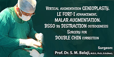
Vertical augmentation genioplasty, LeFort I advancement, malar augmentation, BSSO by Distraction Osteogenesis Surgery for Double Chin Correction

Patient who hated his small jaw presents to our hospital This young man from Australia never liked his retruded chin. It caused him to have a double chin. He had always wished to have a more prominent mandible. His quality of life was also affected by this. The patient had enquired all over Europe, but the costs there were prohibitive. Being a medical doctor himself, he researched the Internet for a quality oral surgeon. His Internet search led him straight to our hospital. He got in contact with our hospital manager who arranged for his travel to India. Treatment plan explained to the patient The patient met with Dr SM Balaji who obtained a detailed history from him. He was very particular that he wanted advancement through distractors. This was because he wanted to monitor for himself the changes as the distractors were activated each day. A treatment plan was then formulated and explained to the patient. His double chin would be corrected. He was then scheduled for surgery. Surgical jaw correction for treatment of double chin A rib graft was first harvested from the patient. A Valsalva maneuver demonstrated absence of any perforation into the thoracic cavity. The incision was then closed with sutures. Attention was next turned to the retrognathic mandible. A vestibular incision exposed the anterior mandibular bone. The chin was then placed forwards with a vertical augmentation genioplasty. Two L-shaped four holed plates were then used to fix the bones of the chin. The posterior mandible was then osteotomized for placement of the distractors. Mandibular distractors were then fixed with screws and tested. There was adequate function of the distractors. Bilateral inferior alveolar nerves were carefully protected during the entire procedure. Attention was then turned to the maxilla. Maxillary osteotomy with placement of bone grafts aided distractor placement. Similar distractors were also utilized here. The incisions were then closed with sutures. The distractors were in stable position. 1 mm distraction per day will be next performed until adequate advancement of jaws. The patient recovered from general anesthesia without any complications. The patient expressed his complete satisfaction with the results before discharge.
Single sitting simultaneous unilateral cleft palate and lip repair
A boy from Ladakh with unilateral cleft lip and palate The patient is a 10-month-old boy with unilateral cleft lip and palate deformity. He lives with his parents in Leh. His family is from a pastoral background. A Good Samaritan from Delhi happened on this little boy during a trek in Ladakh. He offered to help the child and the parents accepted his help. The Good Samaritan did extensive Internet research. This was to find the best cleft lip and palate surgeon who could perform a total cleft repair in one sitting. His search led him straight to our hospital. Treatment planning for simultaneous cleft lip and palate repair Dr SM Balaji examined the patient and ordered imaging studies. He explained to the parents that both cleft lip and repair would undergo surgery. He undertook the surgery after detailed presurgical planning. Simultaneous cleft lip and palate repair surgery performed Under general anesthesia, cleft palate repair was first undertaken. Bilateral palatal flaps were first raised based on the greater palatine vessels. The Levator palatine muscles were then detached from their abnormal positions. These were then reattached into normal position like a hammock. A two layer closure was then done. The nasal floor was first closed in a separate layer with the vomerine flap making a reverse knot. Oral layer was then sutured by vertical mattress sutures. The vertical mattress sutures produce a ridge of thick mucoperiosteum. Flaps were then approximated to each other in the midline. This technique repositions the levator muscle in a more favorable position. Greater palatine osteotomy was then done to mobilize the artery. This was from the greater palatine canal. The suction test performed at the end showed good results. Unilateral cleft lip repair was then performed with the modified Millard’s technique. This resulted in a very good lip seal producing good esthetic results. Parents satisfied with very good surgical results The parents expressed their immense gratitude before discharge from the hospital. Surgery Video
Upper jaw Advancement Surgery Unilateral Cleft Hypoplasia – Lefort 1 Advancement Surgery
Patient presents for maxillary advancement surgery This young lady had been born with a unilateral cleft lip and palate. She had undergone cleft lip repair at our hospital at the age of 2 months. Cleft palate repair was later performed at the age of 10 months. After this, she had rhBMP-2 surgery for uniting the two pieces of the maxilla into one single bone. The patient now has a hypoplastic retruded maxilla with anterior crossbite. This had been causing her cosmetic problems with a deficient upper jaw. She wanted to have this corrected through surgery. The patient has also been undergoing fixed orthodontic treatment for cosmetic teeth alignment. Le Fort 1 maxillary osteotomy planned for the patient Dr SM Balaji is a renowned cleft lip and palate patient rehabilitation specialist. He decided to perform a LeFort 1 osteotomy with maxillary advancement for the patient. Complete correction of the patient’s crossbite occlusion Under general anesthesia, a mucogingivoperiosteal flap was first raised in the maxilla. A LeFort 1 osteotomy was then performed. The maxillary bone was then advanced by 2 cm. It was then stabilized in place with four L-shaped four-holed plates. Occlusion was then checked and deemed to be in perfect alignment. The mucogingivoperiosteal flap was then sutured back in place. She would need further fixed orthodontic treatment to perfect her teeth alignment. Postoperative period was uneventful. The patient expressed her happiness at the results of the surgery before discharge.
Microtia – Ear Lobe Correction – Balaji Dental and Craniofacial Hospital, India
Expert correction of microtia done at our hospital Microtia is a congenital malformation of variable severity of the external ear. Correction of this deformity is through reconstruction surgery. Dr SM Balaji is an expert at microtia correction. This staged procedure gives the best results for microtia correction. Stage 2 microtia correction after successful stage 1 microtia surgery The patient is now 14 years old. He has already undergone stage 1 costal cartilage placement under the skin. This had recreated the form of the left pinna. Stage 2 surgery involves lifting up the reconstructed left pinna. This lies flat against the side of the head after the stage 1 surgery. His left ear lobe was also deformed with skin tags. This too needed corrective surgery. He presents now for stage 2 surgery. Lifting up of reconstructed external ear from side of head Under general anesthesia, an incision was first made around the reconstructed pinna. This was then lifted up and stabilized in position with sutures. Attention was then turned to the deformed ear lobe. A skin incision was first made in the ear lobe and adapted to give normal form to the deformed lobe. Sutures were then used to close the incision. The patient expressed his happiness at the results before discharge from the hospital.
